
How Is Pilonidal Sinus Diagnosed? Tests and Examinations Explained
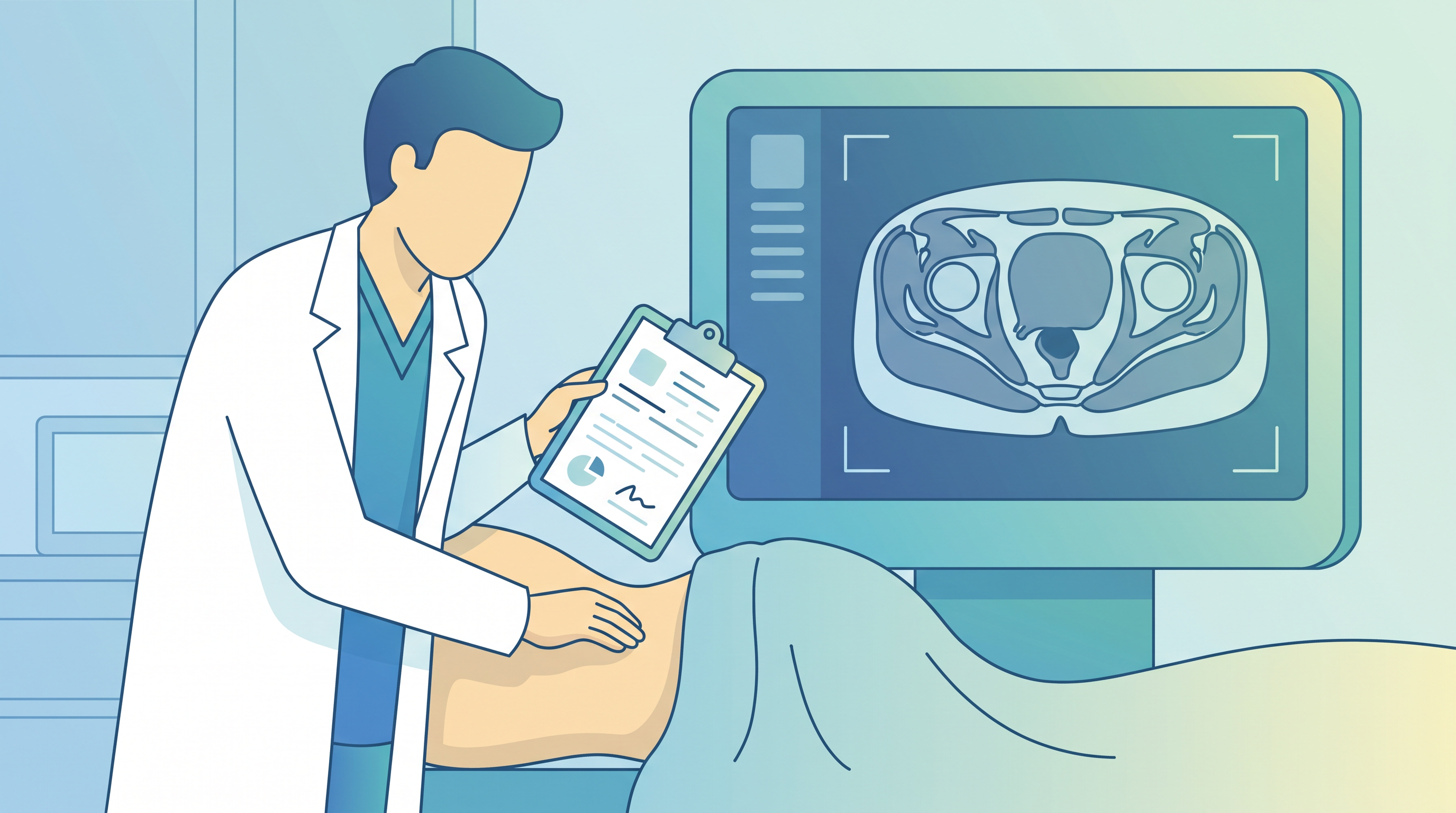
Pilonidal sinus is primarily diagnosed through clinical examination — inspection and palpation of the natal cleft. Ultrasound is used to assess depth and complexity. MRI is reserved for complex or recurrent cases to map all tracts. Blood tests may be ordered if infection is suspected. Most cases are diagnosed without any imaging.
Many patients wonder what to expect when they visit a doctor for pilonidal sinus. The diagnostic process is generally straightforward and involves a clinical examination, sometimes supplemented by imaging. This guide explains exactly what tests and examinations Dr. Adarsh M. Patil uses to diagnose pilonidal sinus disease.
When to Seek Diagnosis
🚨 Seek urgent medical evaluation if you have severe pain, fever, or a rapidly enlarging swelling near your tailbone — do not delay diagnosis in these cases.
Seek urgent care if you have:
- •Severe pain preventing sitting
- •Fever above 38°C
- •Rapidly enlarging tender swelling
- •Significant pus discharge
In Bangalore: Call 108 for emergencies or visit your nearest hospital.
The Diagnostic Process
Diagnosing pilonidal sinus disease involves several steps, starting with a thorough history and physical examination.
Step 1: Medical History
Dr. Adarsh M. Patil at Apollo Clinic Indiranagar, Bangalore will ask about:
- Symptoms: Pain, swelling, discharge, fever
- Duration: How long have you had symptoms?
- Previous episodes: Have you had similar problems before?
- Previous treatment: Any previous surgery or drainage?
- Occupation: Do you sit for long periods?
- Family history: Any family members with pilonidal sinus?
- Medical history: Any conditions affecting healing (diabetes, immune disorders)?
Step 2: Physical Examination
The physical examination is the cornerstone of pilonidal sinus diagnosis.
#What the Examination Involves
1. Inspection: Visual examination of the natal cleft and sacrococcygeal area
- Looking for pits (small openings in the skin)
- Assessing for swelling, redness, or discharge
- Noting the number and location of external openings
2. Palpation: Gentle pressing to assess
- Tenderness and its location
- Extent of swelling
- Whether an abscess is present (fluctuance)
3. Probing: In some cases, a gentle probe is used to assess the depth and direction of the sinus tract
#What Is Found on Examination
| Finding | Significance |
|---------|-------------|
| Single midline pit | Simple pilonidal sinus |
| Multiple pits | Complex multi-tract disease |
| Tender fluctuant swelling | Pilonidal abscess |
| Chronic discharge | Established sinus tract |
| Lateral openings | Complex disease with lateral tracts |
Step 3: Imaging (When Required)
#Ultrasound
When used: For most cases where the depth or complexity is unclear
What it shows:
- Depth of the sinus tract
- Presence of abscess
- Number of tracts
- Proximity to deeper structures
Advantages: Quick, painless, no radiation, widely available
#MRI (Magnetic Resonance Imaging)
When used: Complex or recurrent cases, cases where previous surgery makes anatomy unclear
What it shows:
- Detailed mapping of all sinus tracts
- Relationship to surrounding structures
- Presence of deep infection
Advantages: Most detailed imaging, no radiation
Disadvantages: Expensive, time-consuming, not always necessary
#CT Scan
Rarely used for pilonidal sinus. May be ordered if there is concern about deeper infection or unusual anatomy.
Step 4: Blood Tests
Blood tests are not routinely required for pilonidal sinus diagnosis but may be ordered if:
- Infection is suspected: Full blood count (FBC) to check white blood cell count
- Pre-operative assessment: Before surgery, blood tests assess general health
- Diabetes screening: Diabetes impairs healing and may be detected if not previously diagnosed
What to Expect at Your Consultation
When you visit Dr. Adarsh M. Patil at Apollo Clinic Indiranagar, Bangalore (KMC Reg: 53728, +91 9972446882):
1. You will be asked to lie on an examination table in a comfortable position
2. The examination is brief (5-10 minutes)
3. Dr. Adarsh M. Patil will explain his findings and diagnosis clearly
4. A treatment plan will be discussed and agreed upon
5. Any questions you have will be answered
Differential Diagnosis
Conditions that can mimic pilonidal sinus and are considered during diagnosis:
- Perianal abscess or fistula: Located closer to the anus
- Hidradenitis suppurativa: Chronic skin condition affecting hair follicles
- Sacral dermoid cyst: Congenital cyst near the tailbone
- Furunculosis: Boil caused by skin bacteria
Dr. Adarsh M. Patil's expertise ensures accurate diagnosis and the most appropriate treatment plan.
Dr. Adarsh M. Patil at Apollo Clinic Indiranagar, Bangalore will ask about:
- Symptoms: Pain, swelling, discharge, fever
- Duration: How long have you had symptoms?
- Previous episodes: Have you had similar problems before?
- Previous treatment: Any previous surgery or drainage?
- Occupation: Do you sit for long periods?
- Family history: Any family members with pilonidal sinus?
- Medical history: Any conditions affecting healing (diabetes, immune disorders)?
Step 2: Physical Examination
The physical examination is the cornerstone of pilonidal sinus diagnosis.
#What the Examination Involves
1. Inspection: Visual examination of the natal cleft and sacrococcygeal area
- Looking for pits (small openings in the skin)
- Assessing for swelling, redness, or discharge
- Noting the number and location of external openings
2. Palpation: Gentle pressing to assess
- Tenderness and its location
- Extent of swelling
- Whether an abscess is present (fluctuance)
3. Probing: In some cases, a gentle probe is used to assess the depth and direction of the sinus tract
#What Is Found on Examination
| Finding | Significance |
|---------|-------------|
| Single midline pit | Simple pilonidal sinus |
| Multiple pits | Complex multi-tract disease |
| Tender fluctuant swelling | Pilonidal abscess |
| Chronic discharge | Established sinus tract |
| Lateral openings | Complex disease with lateral tracts |
Step 3: Imaging (When Required)
#Ultrasound
When used: For most cases where the depth or complexity is unclear
What it shows:
- Depth of the sinus tract
- Presence of abscess
- Number of tracts
- Proximity to deeper structures
Advantages: Quick, painless, no radiation, widely available
#MRI (Magnetic Resonance Imaging)
When used: Complex or recurrent cases, cases where previous surgery makes anatomy unclear
What it shows:
- Detailed mapping of all sinus tracts
- Relationship to surrounding structures
- Presence of deep infection
Advantages: Most detailed imaging, no radiation
Disadvantages: Expensive, time-consuming, not always necessary
#CT Scan
Rarely used for pilonidal sinus. May be ordered if there is concern about deeper infection or unusual anatomy.
Step 4: Blood Tests
Blood tests are not routinely required for pilonidal sinus diagnosis but may be ordered if:
- Infection is suspected: Full blood count (FBC) to check white blood cell count
- Pre-operative assessment: Before surgery, blood tests assess general health
- Diabetes screening: Diabetes impairs healing and may be detected if not previously diagnosed
What to Expect at Your Consultation
When you visit Dr. Adarsh M. Patil at Apollo Clinic Indiranagar, Bangalore (KMC Reg: 53728, +91 9972446882):
1. You will be asked to lie on an examination table in a comfortable position
2. The examination is brief (5-10 minutes)
3. Dr. Adarsh M. Patil will explain his findings and diagnosis clearly
4. A treatment plan will be discussed and agreed upon
5. Any questions you have will be answered
Differential Diagnosis
Conditions that can mimic pilonidal sinus and are considered during diagnosis:
- Perianal abscess or fistula: Located closer to the anus
- Hidradenitis suppurativa: Chronic skin condition affecting hair follicles
- Sacral dermoid cyst: Congenital cyst near the tailbone
- Furunculosis: Boil caused by skin bacteria
Dr. Adarsh M. Patil's expertise ensures accurate diagnosis and the most appropriate treatment plan.
1. Inspection: Visual examination of the natal cleft and sacrococcygeal area
- Looking for pits (small openings in the skin)
- Assessing for swelling, redness, or discharge
- Noting the number and location of external openings
2. Palpation: Gentle pressing to assess
- Tenderness and its location
- Extent of swelling
- Whether an abscess is present (fluctuance)
3. Probing: In some cases, a gentle probe is used to assess the depth and direction of the sinus tract
#
What Is Found on Examination
| Finding | Significance |
|---------|-------------|
| Single midline pit | Simple pilonidal sinus |
| Multiple pits | Complex multi-tract disease |
| Tender fluctuant swelling | Pilonidal abscess |
| Chronic discharge | Established sinus tract |
| Lateral openings | Complex disease with lateral tracts |
Step 3: Imaging (When Required)
#Ultrasound
When used: For most cases where the depth or complexity is unclear
What it shows:
- Depth of the sinus tract
- Presence of abscess
- Number of tracts
- Proximity to deeper structures
Advantages: Quick, painless, no radiation, widely available
#MRI (Magnetic Resonance Imaging)
When used: Complex or recurrent cases, cases where previous surgery makes anatomy unclear
What it shows:
- Detailed mapping of all sinus tracts
- Relationship to surrounding structures
- Presence of deep infection
Advantages: Most detailed imaging, no radiation
Disadvantages: Expensive, time-consuming, not always necessary
#CT Scan
Rarely used for pilonidal sinus. May be ordered if there is concern about deeper infection or unusual anatomy.
Step 4: Blood Tests
Blood tests are not routinely required for pilonidal sinus diagnosis but may be ordered if:
- Infection is suspected: Full blood count (FBC) to check white blood cell count
- Pre-operative assessment: Before surgery, blood tests assess general health
- Diabetes screening: Diabetes impairs healing and may be detected if not previously diagnosed
What to Expect at Your Consultation
When you visit Dr. Adarsh M. Patil at Apollo Clinic Indiranagar, Bangalore (KMC Reg: 53728, +91 9972446882):
1. You will be asked to lie on an examination table in a comfortable position
2. The examination is brief (5-10 minutes)
3. Dr. Adarsh M. Patil will explain his findings and diagnosis clearly
4. A treatment plan will be discussed and agreed upon
5. Any questions you have will be answered
Differential Diagnosis
Conditions that can mimic pilonidal sinus and are considered during diagnosis:
- Perianal abscess or fistula: Located closer to the anus
- Hidradenitis suppurativa: Chronic skin condition affecting hair follicles
- Sacral dermoid cyst: Congenital cyst near the tailbone
- Furunculosis: Boil caused by skin bacteria
Dr. Adarsh M. Patil's expertise ensures accurate diagnosis and the most appropriate treatment plan.
#
Ultrasound
When used: For most cases where the depth or complexity is unclear
What it shows:
- Depth of the sinus tract
- Presence of abscess
- Number of tracts
- Proximity to deeper structures
Advantages: Quick, painless, no radiation, widely available
#MRI (Magnetic Resonance Imaging)
When used: Complex or recurrent cases, cases where previous surgery makes anatomy unclear
What it shows:
- Detailed mapping of all sinus tracts
- Relationship to surrounding structures
- Presence of deep infection
Advantages: Most detailed imaging, no radiation
Disadvantages: Expensive, time-consuming, not always necessary
#CT Scan
Rarely used for pilonidal sinus. May be ordered if there is concern about deeper infection or unusual anatomy.
Step 4: Blood Tests
Blood tests are not routinely required for pilonidal sinus diagnosis but may be ordered if:
- Infection is suspected: Full blood count (FBC) to check white blood cell count
- Pre-operative assessment: Before surgery, blood tests assess general health
- Diabetes screening: Diabetes impairs healing and may be detected if not previously diagnosed
What to Expect at Your Consultation
When you visit Dr. Adarsh M. Patil at Apollo Clinic Indiranagar, Bangalore (KMC Reg: 53728, +91 9972446882):
1. You will be asked to lie on an examination table in a comfortable position
2. The examination is brief (5-10 minutes)
3. Dr. Adarsh M. Patil will explain his findings and diagnosis clearly
4. A treatment plan will be discussed and agreed upon
5. Any questions you have will be answered
Differential Diagnosis
Conditions that can mimic pilonidal sinus and are considered during diagnosis:
- Perianal abscess or fistula: Located closer to the anus
- Hidradenitis suppurativa: Chronic skin condition affecting hair follicles
- Sacral dermoid cyst: Congenital cyst near the tailbone
- Furunculosis: Boil caused by skin bacteria
Dr. Adarsh M. Patil's expertise ensures accurate diagnosis and the most appropriate treatment plan.
When used: Complex or recurrent cases, cases where previous surgery makes anatomy unclear
What it shows:
- Detailed mapping of all sinus tracts
- Relationship to surrounding structures
- Presence of deep infection
Advantages: Most detailed imaging, no radiation
Disadvantages: Expensive, time-consuming, not always necessary
#
CT Scan
Rarely used for pilonidal sinus. May be ordered if there is concern about deeper infection or unusual anatomy.
Step 4: Blood Tests
Blood tests are not routinely required for pilonidal sinus diagnosis but may be ordered if:
- Infection is suspected: Full blood count (FBC) to check white blood cell count
- Pre-operative assessment: Before surgery, blood tests assess general health
- Diabetes screening: Diabetes impairs healing and may be detected if not previously diagnosed
What to Expect at Your Consultation
When you visit Dr. Adarsh M. Patil at Apollo Clinic Indiranagar, Bangalore (KMC Reg: 53728, +91 9972446882):
1. You will be asked to lie on an examination table in a comfortable position
2. The examination is brief (5-10 minutes)
3. Dr. Adarsh M. Patil will explain his findings and diagnosis clearly
4. A treatment plan will be discussed and agreed upon
5. Any questions you have will be answered
Differential Diagnosis
Conditions that can mimic pilonidal sinus and are considered during diagnosis:
- Perianal abscess or fistula: Located closer to the anus
- Hidradenitis suppurativa: Chronic skin condition affecting hair follicles
- Sacral dermoid cyst: Congenital cyst near the tailbone
- Furunculosis: Boil caused by skin bacteria
Dr. Adarsh M. Patil's expertise ensures accurate diagnosis and the most appropriate treatment plan.
Blood tests are not routinely required for pilonidal sinus diagnosis but may be ordered if:
- Infection is suspected: Full blood count (FBC) to check white blood cell count
- Pre-operative assessment: Before surgery, blood tests assess general health
- Diabetes screening: Diabetes impairs healing and may be detected if not previously diagnosed
What to Expect at Your Consultation
When you visit Dr. Adarsh M. Patil at Apollo Clinic Indiranagar, Bangalore (KMC Reg: 53728, +91 9972446882):
1. You will be asked to lie on an examination table in a comfortable position
2. The examination is brief (5-10 minutes)
3. Dr. Adarsh M. Patil will explain his findings and diagnosis clearly
4. A treatment plan will be discussed and agreed upon
5. Any questions you have will be answered
Differential Diagnosis
Conditions that can mimic pilonidal sinus and are considered during diagnosis:
- Perianal abscess or fistula: Located closer to the anus
- Hidradenitis suppurativa: Chronic skin condition affecting hair follicles
- Sacral dermoid cyst: Congenital cyst near the tailbone
- Furunculosis: Boil caused by skin bacteria
Dr. Adarsh M. Patil's expertise ensures accurate diagnosis and the most appropriate treatment plan.
Conditions that can mimic pilonidal sinus and are considered during diagnosis:
- Perianal abscess or fistula: Located closer to the anus
- Hidradenitis suppurativa: Chronic skin condition affecting hair follicles
- Sacral dermoid cyst: Congenital cyst near the tailbone
- Furunculosis: Boil caused by skin bacteria
Dr. Adarsh M. Patil's expertise ensures accurate diagnosis and the most appropriate treatment plan.
Medical References & Sources
- [1]Pilonidal sinus diagnosis and management. BMJ, 2022. View source ↗
- [2]Pilonidal disease clinical features. Mayo Clinic, 2023. View source ↗
Dr. Adarsh M. Patil
MS, FMAS — Pilonidal Sinus Specialist, Bangalore
Consult Dr. Adarsh M. Patil — Pilonidal Sinus Specialist
Discuss laser and surgical options, likely recovery, costs, and next steps after an individual assessment.